Pathogenesis, manifestations, and emerging treatments
- Chronic traumatic encephalopathy (CTE) is a neurodegenerative disorder associated with repetitive head injuries.
- Symptoms include a range of psychiatric and psychomotor symptoms, including depression, anxiety, violent behavior, parkinsonism, and visual-spatial difficulties.
- No disease-modifying treatments exist for CTE, and diagnosis can occur only postmortem with a brain biopsy that looks for specific pathology.
- Explain potential causes of chronic traumatic encephalopathy (CTE).
- Describe pharmacologic and nonpharmacologic approaches to the care of a patient with CTE and traumatic brain injury.
- Identify resources to support patients and families.
No relevant financial relationships were identified for any individuals with the ability to control content of the activity.
Expiration: 5/1/27
Chronic traumatic encephalopathy (CTE) is a complex, progressive neurodegenerative disorder associated with repetitive traumatic brain injury (TBI), causes of which include falls and motor vehicle accidents. However, individuals who engage in high-impact sports and military veterans exposed to blast injuries are most vulnerable to TBI and at highest risk for CTE. (See Facts and figures.)
What your patient would like you to know about TBI
Reducing intracranial pressure in patients with traumatic brain injury
Identify the vessel, recognize the stroke
Facts and figures
Confirmation of chronic traumatic encephalopathy (CTE) can occur only after death, so estimating the number of affected individuals has proven difficult. In addition, most CTE prevalence studies tend to focus only on populations most likely to sustain a traumatic brain injury (TBI), such as athletes and military veterans, which limits the ability to determine accurate numbers.
- A postmortem study by LeClair and colleagues of 290 football players from various levels of play revealed that 9.4% (n=51) of high school football players, 47% (n=60) of college football players, and 70.6% (n=166) of players overall had CTE.
- Bieniek and colleagues examined postmortem brain tissue samples in those with a history of CTE manifestations. The study, which included 750 athletes and nonathletes, concluded that 5.6% (42) had CTE pathology or manifestations. The researchers noted the highest rates in those who played American football.
The best estimates of CTE risk have been derived from evaluating the number of those who sustained a TBI.
- According to the U.S. Department of Veteran Affairs, between 2000 and 2019, over 414,000 U.S. military service members have sustained a TBI. The data, however, only included severe TBIs, so actual cases may be higher.
- In 2010, the Centers for Disease Control and Prevention estimated that TBI accounted for approximately 2.5 million emergency department visits in the United States. This represents a potentially significant number of individuals at risk for CTE.
Some researchers have highlighted victims of domestic violence as a population likely at high risk for TBI.
- In a study of 115 participants with a history of experiencing domestic abuse, Zieman and colleagues found that 88% (n=101) experienced multiple brain injuries, and almost 81% (n=93) experienced loss of consciousness. Limitations of research related to CTE and domestic violence include reliance on retrospective analysis.
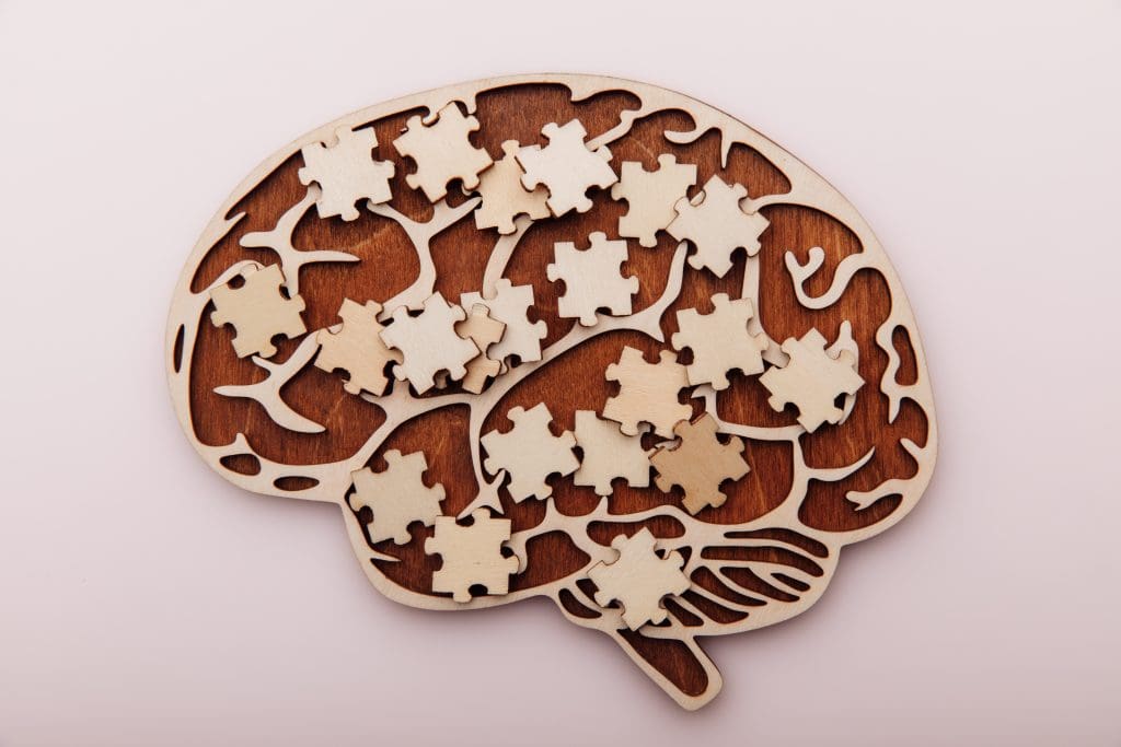
Nurses play a vital role in recognizing those at risk for or suffering from CTE manifestations. Recognizing the associated symptoms in those with a history of TBI is the first step in empowering individuals to find answers and heal. Understanding CTE pathogenesis, manifestations, and emerging therapies will help nurses provide quality care to patients and their families.
Pathogenesis
CTE occurs primarily as a result of microglia neuronal cell-mediated kinase activation resulting from head injury. This activation results in abnormal phosphorylation (hyperphosphorylation) of tau protein, which leads to the development of pathologic neurofibrillary tangles, tau protein deposits, and eventual neuronal death. Normally, microglia and tau proteins exist throughout the central nervous system and have neuroprotective properties.
Microglial dysfunction may play a key role in CTE formation. Mild-to-severe TBI can disrupt the blood–brain barrier, resulting in the proliferation of toxic levels of neurochemicals into the parenchyma. The microglia become primed, and if they remain primed during a subsequent incident of head trauma, they can release further toxic levels of pro-inflammatory cytokines. With repetitive head trauma, the microglia remain overstimulated and can’t switch back to a neuro-reparative mode. These insults lead to continued neuro-inflammation (via kinase-phosphate imbalances), tissue injury, and chronic neurodegeneration.
Tau protein plays an important role in stabilizing microtubule fibrils by binding tubulin. Tau strengthens the cytoskeleton of the neuron, which facilitates the neurite to branch out and eventually become axons or dendrites. The imbalance between kinase and phosphatase activity causes the phosphorylation of tau, which leads to tubulin separation. The separation leads to a dysfunction of microtubules and tau becoming insoluble, resulting in an accumulation of tau (tau oligomers) in neuron bodies. This accumulation leads to DNA damage and cytoskeletal/synaptic dysfunction. As a protective measure, neurofibrillary tangle formations are created to capture and hold the tau oligomers. However, these protective tangles ultimately lead to neuronal death.
Rapid acceleration, deceleration, or rotation forces to the brain can lead to other pathological changes such as diffuse axonal injury. The elongating forces acting on axonal membranes increase their permeability. The following influx of calcium leads to a cascade of events that promote the degradation of the axon cytoskeletal network. Axonal injury caused by the shearing force of head trauma contributes to neuronal death. Changes in the limbic system resulting from CTE include atrophy of the hippocampus, entorhinal cortex, and amygdala.
The underlying changes in the brain can contribute to the development of psychiatric illness and, perhaps, the onset or acceleration of pathologically similar neurodegenerative diseases such as Alzheimer’s disease (AD), frontotemporal lobar degeneration, and Lewy body dementia. These neurodegenerative diseases also are associated with tau accumulation; however, growing evidence recognizes that the tau buildup in CTE may be distinct from these other disorders.
Clinical manifestations
CTE can arise from multiple hits to the head, even if it doesn’t lead to loss of consciousness or concussive symptoms. Although CTE is thought to occur as a result of repeated injuries, more research will help determine the number and types of injuries associated with CTE development. Manifestations can occur years after the injuries.
CTE progresses through four stages, each marked by distinct neurological, psychiatric, and motor manifestations, most of which involve changes in cognition, behavior, or mood. Patients with typically mild Stage 1 CTE experience short-term memory loss, difficulty concentrating, depression, mood swings, aggression, suicidal thoughts, and headaches. In Stage 2, emotional and mood disturbances, including depression and suicidal thoughts, continue. Explosivity, outbursts, and language challenges (dysarthria) also may occur. Stage 3 is marked by aggression, apathy, and increasing memory loss. In this stage, individuals may have visual–spatial deficits, including difficulty with sense of direction or estimating an object’s size or shape. Lack of spatial awareness can manifest as unawareness of oneself or an object in space. These deficits can cause issues with following directions, reading a map, or doing a puzzle. In Stage 4, memory loss becomes severe and individuals may exhibit paranoid behavior. Parkinsonism—which includes uncontrollable or unintentional movement, as well as gait, balance, and coordination issues—may occur. Cognitive abilities continue to worsen, eventually impairing the ability to read, think, learn, and reason. (See CTE stages).
CTE stages
The progression of chronic traumatic encephalopathy (CTE) occurs in four stages with increasing manifestations.

Diagnosis
CTE diagnosis can occur only postmortem with a brain biopsy that looks for specific pathology. Postmortem analysis includes evaluation for perivascular accumulations of hyperphosphorylated tau in neurons concentrated deep into the surface cortical sulci. In contrast, AD lesions are deeper and distributed in cortical areas. CTE is staged according to the extent/severity of the tau pathology.
Suspicion of CTE is based on a history of TBI and a thorough neurological and neuropsychological assessment. The neurologic exam includes evaluation of cranial nerves, motor and sensory function, balance, coordination, and reflexes. Mental status includes appearance, alertness, speech, behavior, mood, affect, thought process and content, memory, and judgment and reasoning. (See Screening and assessment tools.)
Screening and assessment tools
Several tools can aid screening and assessment of individuals suspected of having chronic traumatic encephalopathy.
Mental status evaluation
- Mini Mental Status Examination
(cgatoolkit.ca/Uploads/ContentDocuments/MMSE.pdf) - Montreal Cognitive Assessment
(mocacognition.com)
Depression
- Beck Depression Inventory
- Patient Health Questionnaire-2 (PHQ-2)
(cde.nida.nih.gov/sites/nida_cde/files/PatientHealthQuestionnaire-2_v1.0_2014Jul2.pdf) - If the PHQ-2 is positive, follow up with the Patient Health Questionnaire-9
(apa.org/depression-guideline/patient-health-questionnaire.pdf).
Anxiety
- Beck Anxiety Inventory
(theinvictusclinic.com/wp-content/uploads/2019/10/Beck-Anxiety-Inventory.pdf) - General Anxiety Disorder-7 (GAD-7) (adaa.org/sites/default/files/GAD-7_Anxiety-updated_0.pdf).
GAD-7 also is useful in screening for post-traumatic stress disorder, panic disorder, and social anxiety disorder.
Proposed clinical diagnostic criteria for CTE (traumatic encephalopathy syndrome) include a history of multiple impacts, absence of comorbid disease that could account for the patient’s symptoms, presence of symptoms for at least 12 months, at least one core clinical feature (behavioral, mood, or cognitive impairment), and supportive features such as decline over 12 months or longer, impulsivity, and headaches.
Brain imaging with computerized tomography or magnetic resonance imaging (MRI) can help rule out other conditions. Preliminary results of early research that focused on using MRI to detect structural atrophies specifically associated with CTE offer hope for acquiring an in vivo diagnostic tool. Using serum biomarkers to diagnose CTE has proven difficult because of the similar pathology with AD. However, emerging research focused on assessing multiple biomarkers, such as total-tau, soluble triggering receptor expressed on myeloid 2, and neurofilament light chain, may indicate microglia activation, neuroinflammation, and axonal injury. Although results look promising, the medical community has yet to agree on how to incorporate the new data into the diagnostic criteria.
Treatment
No disease-modifying treatments exist for CTE. Currently, treatment strategies are similar to those for TBI or other neurodegenerative disorders such as Parkinson’s disease. Pharmacologic and nonpharmacologic treatment offers supportive care and focuses on managing clinical symptoms as they arise.
Pharmacologic management
Standard pharmacologic management includes selective serotonin reuptake inhibitors (SSRIs) (such as fluoxetine and citalopram), atypical antipsychotic medication (such as aripiprazole and clozapine), and cholinesterase inhibitors (such as donepezil). Dopamine agonists (such as carbidopa/levodopa and memantine) can help manage cognitive, motor, and mood-related symptoms. Recent research suggests that sertraline is most effective at treating TBI-related depression. However, only limited research exists regarding SSRI effectiveness in patients after a TBI; additional studies are needed.
Nonpharmacologic management
Nonpharmacologic TBI management includes various cognitive rehabilitation therapies (CRTs), which aim to enhance function and independence based on individual needs and symptoms. Restorative CRT works to strengthen or regain a patient’s lost function via memory tests and attention-span training. Compensatory CRT uses cueing, memory tool devices, and calendars to help patients work around their deficits. Mood and behavior therapy, such as cognitive behavioral therapy and psychodynamic therapy, can facilitate anxiety and depression management. In addition, mindfulness can help patients remain aware of their present state and avoid becoming overly reactive and overwhelmed.
Physical therapy, aerobic exercise, and nutrition also can help manage symptoms. For example, vestibular and ocular rehabilitation may benefit those with vestibular inner ear or visual symptoms. Also, the Mediterranean diet may mitigate microglia-mediated neuroinflammation. Although no single definition or consensus of the Mediterranean diet exists, the key characteristics include high consumption of fruits, vegetables, whole grains (for example, whole wheat bread and brown rice), nuts, legumes (for example, lentils, kidney beans, and black beans), and nuts/seeds (for example, almonds, walnuts, and pistachios). Extra virgin olive oil serves as the principal source of fat in this diet, which also includes fish rich in omega-3 fatty acids such as herring, salmon, and sardines. (See Prevention.)
Prevention
The only way to prevent chronic traumatic encephalopathy is to prevent traumatic brain injury.
- Use safety equipment, such as helmets, when participating in sports, and seatbelts, when driving.
- Limit the use of alcohol and drugs, especially when driving or engaging in sports.
- Use window locks and gates at the top of stairs to reduce falls in children.
- Install guard rails, remove throw rugs, and ensure proper lighting to reduce falls in older adults. In addition, older adults should engage in strength and balance training and be aware of medications that can cause hypotension or sedation.
Emerging treatments
Pharmacologic therapies under investigation for TBI and CTE prevention include neurosteroids, kinase inhibitors, immunotherapy with monoclonal antibodies, anti-inflammatory therapy with omega-3 fatty acids, and hyperbaric oxygen therapy (HBOT).
Neurosteroids. Estrogen, testosterone, and progesterone aid neuroinflammation prevention. Ripley and colleagues reported that up to 80% of men who suffered a severe TBI experienced hypogonadism and low levels of testosterone. Symptoms associated with hypogonadism, such as fatigue, irritability, and depression, also have been found in CTE. Hormone regulation as part of post-TBI symptom management is being evaluated and incorporated in treatment protocols.
Lithium, a kinase inhibitor that targets tau phosphorylation, is used to treat bipolar disorder. It addresses symptoms such as depression, labile mood, and suicidal ideation, which can occur in patients with CTE. Preclinical rodent studies indicate that lithium can slow the progression of CTE by blocking kinase activity, preventing tau uptake, preserving the blood–brain barrier, and reducing inflammation. Other drugs under investigation that also target tau phosphorylation include I.V. simvastatin, roscovitine, and GSK3 inhibitors.
Immunotherapy. Other research focuses on immunotherapy using monoclonal antibodies and inflammatory drugs (such as calpain-2 inhibitors). Preclinical studies have shown a significant reduction in the accumulation of tau and pro-inflammatory cytokines.
Anti-inflammatory therapy. Use of omega-3 fatty acids post-TBI has shown long-term tissue restoration and cognitive recovery in rodent brains. The use of omega-3 polyunsaturated fatty acids before and after TBI resulted in decreased learning and memory deficits. Currently, no researchers are conducting human trials to test the effectiveness of omega-3 fatty acids on TBI recovery. However, Heileson and colleagues found a reduction in serum neurofilament light chain in members of a college football team who took high doses of omega-3 fatty acids compared to a control team. Interestingly, the control group experienced a significant increase in serum neurofilament light chain.
Lewis and colleagues suggest that diffuse axonal injury severity can be mitigated with something as simple as high doses of omega-3s. These fatty acids may increase the neuroprotection of brain cells and reduce neuroinflammation and neurodegeneration. Researchers continue to investigate the use of omega-3 fatty acids in preventing and treating TBI. As no serious side effects have been reported, some providers have incorporated its use into their treatment protocols.
HBOT. Harch and colleagues reported that HBOT can increase healing in microscopic cerebral wounds caused by TBI. The therapy may increase cognition and mood while reducing headaches and symptoms associated with post-traumatic stress disorder, which can occur after a TBI.
Uncovering mysteries
CTE, an insidious disease, slowly leads to extensive psychiatric and motor symptoms and significantly impaired quality of life. Knowledge of a patient’s TBI history can aid identification of the underlying cause of presenting symptoms, support well-informed decisions, and ensure patients and families receive proper education. The mysteries embedded in the pathology of CTE offer a range of research possibilities involving medical and psychiatric treatment.
Resources
The following traumatic brain injury (TBI) resources can help patients and families connect with support groups, counseling, and treatment.
- Brain Care Concussion Clinic (braincare.center) developed omega-3 protocols, which can be used as prophylactic and post-injury measures to potentially reduce the negative effects of TBI.
- Brain Health Education and Research Institute (brainhealtheducation.org) focuses on brain health research, education, and treatment. Treatment protocols include the use of neurosteroids, hormone therapy, specialized diets, omega-3, and other nutraceuticals. The institute specializes in treating veterans and first responders in addition to the general population.
- Brain Injury Association of America (biausa.org) promotes research, advocacy, and education to improve the care and treatment of patients with TBI. In addition, this nonprofit connects families and patients with support groups and counseling services.
- Concussion Legacy Foundation (concussionfoundation.org) focuses on sports-related concussions and works to advance research, education, and policy to improve safety in sports and other activities that can cause TBI. The nonprofit group also offers counseling, resources, and support groups for patients, families, and caregivers impacted by sports-related TBI.
- Make the Connection (maketheconnection.net) provides links to support groups and resources for education, counseling, and treatment. The organization also assists with locating nearby Veterans Affairs resources.
- Millennium Health Centers (tbimedlegal.com) offer nutraceuticals to reduce neuroinflammation and provide hormone regulation if needed. The centers also offer in-person and online a TBI diagnosis and treatment lecture series to healthcare providers.
- TBI Help Now (tbihelpnow.org) focuses on resolving TBI by healing the brain at the root cause.
Jahaziel Marrero is a firefighter and paramedic with Miami-Dade Fire Rescue attached to the Miami Dade Police Department’s Crisis Response Unit in Miami, Florida. Lucie Dlugasch is a clinical professor at Florida International University in Miami.
American Nurse Journal. 2024; 19(5). Doi: 10.51256/ANJ052406
References
Adams JW, Alvarez VE, Mez J, et al. Lewy body pathology and chronic traumatic encephalopathy associated with contact sports. J Neuropathol Exp Neurol. 2018;77(9):757-68. doi:10.1093/jnen/nly065
Alosco ML, Mian AZ, Buch K, et al. Structural MRI profiles and tau correlates of atrophy in autopsy-confirmed CTE. Alzheimers Res Ther. 2021;13(1):193. doi:10.1186/s13195-021-00928-y
Bieniek KF, Blessing MM, Heckman MG, et al. Association between contact sports participation and chronic traumatic encephalopathy: A retrospective cohort study. Brain Pathol. 2020;30(1):63-74. doi:10.1111/bpa.12757
Brain Health Education and Research Institute.Omega-3 protocol for brain injury. brainhealtheducation.org/omega-3-protocol
Breen PW, Krishnan V. Recent preclinical insights into the treatment of chronic traumatic encephalopathy. Front Neurosci. 2020;14:616. doi:10.3389/fnins.2020.00616
Centers for Disease Control and Prevention. Get the facts about TBI. April 20, 2023. cdc.gov/traumaticbraininjury/get_the_facts.html
Centers for Disease Control and Prevention. Traumatic brain injury & concussion: Prevention. May 12, 2021. cdc.gov/traumaticbraininjury/prevention.html
CTE Society. Going through the stages of CTE. 2020. ctesociety.org/latest_news/going-through-the-stages-of-cte
Franz CK, Joshi D, Daley EL, et al. impact of traumatic brain injury on amyotrophic lateral sclerosis: From bedside to bench. J Neurophysiol. 2019;122(3):1174–85. doi:10.1152/jn.00572.2018
Harch PG, Andrews SR, Rowe CJ, et al. Hyperbaric oxygen therapy for mild traumatic brain injury persistent postconcussion syndrome: A randomized controlled trial. Med Gas Res. 2020;10(1):8-20, doi:10.4103/2045-9912.279978
Heileson JL, Anzalone AJ, Carbuhn AF, et al. The effect of omega-3 fatty acids on a biomarker of head trauma in NCAA football athletes: A multi-site, non-randomized study. J Int Soc Sports Nutr. 2021;18(1):65. doi:10.1186/s12970-021-00461-1
Hu P, Lu Y, Pan BX, Zhang WH. New insights into the pivotal role of the amygdala in inflammation-related depression and anxiety disorder. Int J Mol Sci. 2022;23(19):11076. doi:10.3390/ijms231911076
Katsumoto A, Takeuchi H, Tanaka F. Tau pathology in chronic traumatic encephalopathy and Alzheimer’s disease: Similarities and differences. Front Neurol. 2019;10:980. doi:10.3389/fneur.2019.00980
LeClair J, Weuve J, Fox MP, et al. Relationship between level of American football playing and diagnosis of chronic traumatic encephalopathy in a selection bias analysis. Am J Epidemiol. 2022;191(8):1429-43. doi:10.1093/aje/kwac075
Lewis MD. When Brains Collide: What Every Athlete and Parent Should Know About the Prevention and Treatment of Concussions and Head Injuries. Austin, TX: Lioncrest Publishing; 2016.
Li W, Risacher SL, McAllister TW, Saykin AJ. Traumatic brain injury and age at onset of cognitive impairment in older adults. J Neurol. 2016;263(7):1280-5. doi:10.1007/s00415-016-8093-4
Lucke-Wold BP, Logsdon AF, Nguyen L, et al. Supplements, nutrition, and alternative therapies for the treatment of traumatic brain injury. Nutr Neurosci. 2018;21(2):79-91. doi:10.1080/1028415X.2016.1236174
McCambridge M, Stinson MJ. Advances in chronic traumatic encephalopathy. JAAPA. 2020;33(4):39-42. doi:10.1097/01.JAA.0000654008.48462.01
Narapareddy BR, Narapareddy L, Lin A, et al. Treatment of depression after traumatic brain injury: A systematic review focused on pharmacological and neuromodulatory interventions. Psychosomatics. 2020;61(5):481-97. doi:10.1016/j.psym.2020.04.012
Pierre K, Dyson K, Dagra A, Williams E, Porche K, Lucke-Wold B. Chronic traumatic encephalopathy: Update on current clinical diagnosis and management. Biomedicines. 2021;9(4):415. doi:10.3390/biomedicines9040415
Ripley DL, Gerber D, Pretz C, Weintraub AH, Wierman ME. Testosterone replacement in hypogonadal men during inpatient rehabilitation following traumatic brain injury: Results from a double-blind, placebo-controlled clinical pilot study. NeuroRehabilitation. 2020;46(3):355-68. doi:10.3233/NRE-192992
Scrimgeour, AG, Condlin ML, Loban A, DeMar JC. Omega-3 fatty acids and vitamin D decrease plasma T-Tau, GFAP, and UCH-L1 in experimental traumatic brain injury. Front Nutr. 2021;8:685220. doi:10.3389/fnut.2021.685220
Tagge CA, Fisher AM, Minaeva OV, et al. Concussion, microvascular injury, and early tauopathy in young athletes after impact head injury and an impact concussion mouse model. Brain. 2018;141(2):422-58. doi:10.1093/brain/awx350
UCSF Weill Institute for Neurosciences. A health care provider’s guide to chronic traumatic encephalopathy (CTE). memory.ucsf.edu/sites/memory.ucsf.edu/files/wysiwyg/UCSF_CTE_Providers_7-13-17.pdf
UCSF Weill Institute for Neurosciences. Memory and Aging Center. Chronic traumatic encephalopathy. memory.ucsf.edu/dementia/chronic-traumatic-encephalopathy
U.S. Department of Veteran Affairs. Traumatic brain injury (TBI). research.va.gov/topics/tbi.cfm
Vile AR, Atkinson L. Chronic traumatic encephalopathy: The cellular sequela to repetitive brain injury. J Clin Neurosci. 2017;41:24-9. doi:10.1016/j.jocn.2017.03.035
Wu Y, Zhang J, Feng X, Wei J. Omega-3 polyunsaturated fatty acids alleviate early brain injury after traumatic brain injury by inhibiting neuroinflammation and necroptosis. Transl Neurosci. 2023;14(1): 20220277. doi:10.1515/tnsci-2022-0277
Xie Z, Meng J, Wu Z, et al. The dual nature of microglia in Alzheimer’s disease: A microglia-neuron crosstalk perspective. Neuroscientist. 2022;29(5):616-38 doi:10.1177/10738584211070273
Yue JK, Burke JF, Upadhyayula PS, et al. Selective serotonin reuptake inhibitors for treating neurocognitive and neuropsychiatric disorders following traumatic brain injury: An evaluation of current evidence. Brain Sci. 2017;7(8):93. doi:10.3390/brainsci7080093
Zieman G, Bridwell A, Cárdenas JF. Traumatic brain injury in domestic violence victims: A retrospective study at the Barrow Neurological Institute. J Neurotrauma. 2017;34(4):876-80 doi:10.1089/neu.2016.4579
Key words: chronic traumatic encephalopathy, traumatic brain injury, CTE, TBI, neurodegenerative disorders


1 Comment. Leave new
course very informative