Prevent complications and provide education.
- For the foot to properly perform its main functions of weight bearing and propulsion, 26 bones, 33 joints, and over 100 tendons, muscles, ligaments, blood vessels, nerves, and nails must work together.
- A 3-minute foot assessment screens for lower extremity complications by providing high impact, critical information on the patients’ lower extremity health.
- Foot assessment provides an opportunity for early problem identification and can prevent long-term impact to physical function and quality of life.
For the foot to properly perform its main functions of weight bearing and propulsion, 26 bones, 33 joints, and over 100 tendons, muscles, ligaments, blood vessels, nerves, and nails must work together. Given the complexity of the foot, it should come as no surprise that Bryant and Nix reported that 75% of Americans experience foot problems. In addition, foot problems impact 71% to 87% of the elderly, frequently as a result of functional deficits in vision or dexterity.
Diabetic foot ulcers in ambulatory settings
Nurses and preventive foot care
Complications of foot disease affect functional mobility and can increase the risk of falls. As we age, the epidermis and dermis thin, placing the foot at increased risk for pain and wounds. The comorbid conditions we amass as we age that affect foot health include vascular-related trophic changes and edema. A significant positive correlation exists between foot health and quality of life. This largely results from how foot problems affect mobility, vitality, and social interaction. Identifying issues early via a thorough foot assessment can prevent long-term impact to physical function and quality of life.
3-minute foot assessment
A simple 3-minute foot assessment can help you screen patients for lower extremity complications. In the outpatient environment, the assessment is recommended at every visit for all patients with a suspected or confirmed diagnosis of comorbidities (such as peripheral arterial disease [PAD] or diabetes) that place them at increased risk for complications of foot disease. This evidence-based tool offers a quick method for early detection. It also includes recommendations for a risk-based referral system to connect the patient with specialist care.
Each minute of the 3-minute foot assessment focuses on a single important facet: ask, look, teach. These high-impact components provide critical information about the patients’ lower extremity health.
Minute 1: Ask
During the first minute of the 3-minute foot assessment, ask about the patient’s medical and social history as well as any future appointments. You want to find out whether they’ve had any previous foot or leg surgeries, amputations, or vascular interventions. Ask the patient if they have an open foot wound. In addition, include questions related to social factors, such as tobacco use and blood glucose control.
Use this minute to ask about the patients’ health status. For example, the presence of burning, tingling, or loss of sensation in the legs or feet may indicate polyneuropathy or restless leg syndrome. These diagnoses carry increased risk of tissue loss and PAD. Leg or foot pain with activity and changes in skin color or skin lesions also provide valuable clues that may signal the presence of claudication or PAD.
Ask who the patient sees for lower extremity care. Common specialties include podiatry, vascular surgery, and orthopedics. Ascertaining the last time the patient saw a specialist and whether they have upcoming appointments can help guide you in recommending appropriate follow-up for any concerns you uncover.
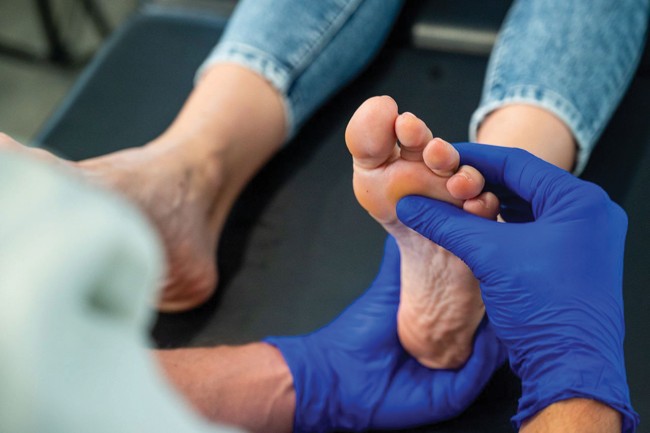
Minute 2: Look
During the second minute of the assessment, perform visual and physical assessments of the musculoskeletal, neurologic, vascular, and dermatologic (including nails) systems.
Musculoskeletal assessment. A brief musculoskeletal assessment includes range of motion in the joints of the ankle and foot. Ask the patient to move the ankle joint in a circular motion, flex the foot, and extend or point the foot. Note any obvious deformities and confirm with the patient how long they’ve existed. A patient with neuropathy who’s midfoot is hot, red, inflamed, or newly deformed may require urgent referral to an appropriate provider to evaluate for complications such as osteomyelitis or Charcot foot. In the presence of neuropathy, these potentially limb-threatening findings can occur with or without pain.
Neurologic assessment. You can accomplish a neurologic assessment by determining the patient’s responsiveness to light touch. The Semmes-Weinstein monofilament test, the gold standard in neurologic foot assessment, involves placing a specific kind of flexible plastic over various anatomical areas of the foot. The patient identifies when they feel the monofilament. If your organization doesn’t have this test, use your hand. The ability to feel light touch (protective sensation) warns the body of potential danger to tissues, including temperature extremes and ill-fitting footwear. Impairment in lower extremity sensory nerves to inform the dorsal root ganglia of dangerous stimuli, which normally triggers activity avoidance, can result in ulcer formation and diminished wound healing.
Vascular assessment. You may not see physical manifestations of vascular disease during a clinical exam, but the prevalence of lower extremity arterial disease in some conditions, such as diabetes, makes it crucial to perform a thorough vascular assessment. Compare the temperature between the calves and feet, palpate the dorsalis pedis and posterior tibial pulses, and note the presence of hair on the lower extremity. Many disease states can contribute to hair loss, but it’s a common finding in PAD due to decreased perfusion of the follicle. (See Peripheral arterial disease.)
Peripheral arterial disease
 Findings typical in peripheral arterial disease include rubor, pallor to the distal fourth digit, and self-avulsed hallux nail plate with moist eschar as a result of lack of perfusion.
Dermatologic assessment. During this part of the assessment, you can uncover foot wounds that many patients, especially those with polyneuropathy, aren’t aware of. Frequently, providers miss wounds encrusted with drainage and eschar because they mistake them as a scab or hyperkeratotic plaque related to poor hygiene. Fissures may be open at the base, and thus a risk for infection. These should be noted during assessment and evaluated thoroughly.
Age-related skin changes that also affect barrier function include declining lipid content and sebum production. The sebaceous glands that produce sebum decrease their production by about 65% as skin ages, although the size and number of glands remain stable. However, eccrine glands (sweat glands) reduce in number by approximately 15%, which contributes to an overall 70% reduction in sweating. These changes contribute to the increased incidence of anhidrosis (dry skin) in older adults.
Autonomic neuropathy, which occurs in patients with diabetes in addition to motor and sensory neuropathies, affects the intact barrier function of the skin. Dysfunction of autonomic nerves leads to anhidrosis. Alterations in moisture content compromise the acid mantle of the skin and lead to cracks and fissures that may become portals of entry for pathogens. Altered skin barrier function leads to deficient defense against pathogenic invasion and contributes to increased rates of skin and soft-tissue infection in patients with diabetes. Dry skin flakes and hyperkeratotic plaques also function as reservoirs for bacteria and fungi, which prevent proper cleansing. (See Autonomic neuropathy.)
Autonomic neuropathy
 Patients with autonomic neuropathy and type 2 diabetes may experience hyperkeratotic plaques (seen here on the left plantar foot of a patient).
Keratinocytes, the skin cells most prevalent in the epidermis and which produce keratin, become phenotypically “switched on” in a number of inflammatory disease states including wound healing, repetitive injury or stress, and obesity. An overproduction of keratin can create hyperkeratotic plaques, hyperkeratosis, keratomas, or callus. (See Hyperkeratosis.)
Hyperkeratosis
 This photo shows the distal left forefoot of a patient with extreme morbid obesity, lymphedema, elongated nails, a dystrophic and discolored hallux nail plate, visible subungual debris, and hyperkeratosis.
A foot callus in a patient at high risk for skin conditions may indicate a repetitive injury. The callus itself increases soft-tissue pressure beneath its firm surface. Callus frequently precedes foot ulceration and abscesses. These flakes, crusts, and plaques protect bacteria and fungus during cleansing and allow them to recolonize surfaces quickly. (See Callus.)
Callus
The following photos show a plantar foot keratoma or callus with blistering to disto-lateral aspect (pre-debridement at left; post-debridement at right).

Commonly encountered onychopathology (altered anatomical and disease states of the toenails) includes paronychia, onychocryptosis, onychogryphosis, and onychomycosis. Paronychium (soft tissue surrounding the nail plate) serves as the origin of the paronychia disease process, which can lead to a painful pustule. The nail matrix originates in the nail root—a tapered anatomic structure that extends 7 mm to 8 mm beneath the eponychium’s (cuticle’s) seal between the nail and the digit. Onychocryptosis (ingrown toenail) refers to the “crypt” or small pit or recess where the nail plate has extended beyond the normal anatomical position. Onychogryphosis presents as large, deformed, hypertrophic nails. The Latin “gryph” (short for gryphon) refers to the nail plate appearing like the talon or claw of a griffin. Nails displaying onychogryphosis sometimes are called “ram’s horn nails” because of untrimmed, thickened nail plates that curl like a ram’s horn. The loss of normal nail translucency during these disease states can increase difficulty in identifying the free nail border, which increases the risk of soft-tissue damage during nail care. (See Paronychia.)
Paronychia
 This photo shows a discolored left hallux nail plate with paronychia and erythema.
Fungal infection affects 20% to 25% of the population with the incidences as high as 40% to 60% in some regions. Onychomycosis refers to fungal infections of the nails. Nail infection is most commonly caused by dermatophytes (tinea unguium), followed by single-celled yeast candida and other non-dermatophyte mould. Dermatophytes, a group of fungi that inhabit the keratin-rich stratum corneum of human skin, can harmlessly colonize humans. Disease occurs in the presence of structural or functional harm. A healthy nail plate that’s properly vascularized appears translucent and pink. Mycotic nails may appear yellow, tan, or brown. The nail plate may be thickened, brittle, and easily break off into small pieces.
Fungal infection that impacts the remainder of the foot is called tinea pedis. The classic presentation of tinea pedis includes pruritus, erythema, and interdigital maceration.
Minute 3: Teach
The final minute of the 3-minute foot assessment provides you with an opportunity to educate the patient about prevention, intervention for any abnormalities, and recommended follow-ups.
Professional organizations and clinical advocacy groups—including the Association for the Advancement of Wound Care, Amputation Prevention Alliance, American Heart Association’s Peripheral Artery Disease National Action Plan, and the American Diabetes Association—endorse several foot care recommendations. Although slight recommendation variations exist among the groups, they agree that healthy foot care includes daily foot inspection, wearing socks and shoes, not walking barefoot, and selecting proper footwear. (See Foot care recommendations.)
Foot care recommendations
Several professional organizations and advocacy groups support the following foot care recommendations:
- Wash feet thoroughly every day.
- Dry them thoroughly, including between the toes.
- Moisturize feet; don’t moisturize between toes.
- Keep toenails trim and use an emery board to file down sharp edges.
- Check feet for sores, cuts, blisters, corns, or redness each day.
Contact provider if any are found.
- Wear moisture-wicking socks.
- Before donning shoes, check for sharp objects (such as small rocks).
- Wear shoes that fit well and don’t rub the feet.
Avoid the following:
- Soaking feet
- Smoking
- Walking barefoot (including inside your house)
If patients can’t visualize the entirety of both feet on their own, encourage them to recruit a friend or family member to help them in a daily foot inspection. In addition, emphasize the importance of seeking prompt medical attention for foot problems, including calluses, minor traumatic injuries, signs or symptoms of infection, and sores. Appropriate footwear refers to shoes that aren’t too small or too large, that don’t rub against a single area of the foot, and aren’t more than a year old or worn out.
Refer patients with diabetes or those seeking further education (such as antalgic [altered gait] training, appropriate footwear, or injury prevention) to a foot care specialist. Anyone with these concerns may benefit from this care, even if they aren’t experiencing loss of protective sensation or PAD. Patients with diabetes generally see a foot care specialists at least once a year to have toenails, calluses, and corns trimmed. The specific time frame is determined based upon patient need.
Specialized competence
Foot assessment provides an opportunity for early identification of issues and can help prevent long-term impact on physical function and quality of life. Access to quality foot care is associated with preventing complications (such as major lower extremity amputation), increasing quality of life, and decreasing pain.
The complexity of the foot and the prevalence of foot problems require specialized competence and professionalism for nurses who provide foot care assessment, intervention, and education in all settings. Certified foot care nurses have elevated patient care in hospitals, long-term care facilities, podiatry offices, and in their own homes. The increased credibility, recognition, and authority of certification has led to greater opportunities to develop foot care interventions that improve patient well-being and enhance risk management. Visit wocncb.org/certification/foot-care-certification to learn more about becoming a certified foot care nurse.
Laura Swoboda is the director of skin and wound care at the Healing Institute in Milwaukee, Wisconsin.
American Nurse Journal. 2024; 19(9). Doi: 10.51256/ANJ092422
References
Almaawi A, Alqarni H, Thallaj AK, et al. Foot health and quality of life among adults in Riyadh, Saudi Arabia: A cross-sectional study. J Orthop Surg Res. 2023;18(1):192. doi:10.1186/s13018-023-03677-w
American Heart Association. Helping Your Patients with Peripheral Artery Disease—Lower Extremity: A Clinician’s Guide. 2023. ahapad.ksw-gtg.com/publication/?m=46 677&i=787993&p=1&ver=html5
Berbudi A, Rahmadika N, Tjahjadi AI, Ruslami R. Type 2 diabetes and its impact on the immune system. Curr Diabetes Rev. 2020;16(5):442-9. doi:10.2174/1573399815666191024085838
Bodman MA, Syed HA, Krishnamurthy K. Onychomycosis. StatPearls. January 9, 2024. ncbi.nlm.nih.gov/books/NBK441853
Bryant RA, Nix DP. Acute & Chronic Wounds: Intraprofessionals From Novice to Expert. 6th ed. Philadelphia, PA: Elsevier; 2023.
Choo ZN, Lipner SR. Onychogryphosis is associated with dermatologic and vascular disease: A case-control study of the All of Us Research Program. Skin Appendage Disord. 2023;9(4):252-7. doi:10.1159/000530096
Conte MS, Bradbury AW, Kolh P, et al. Global vascular guidelines on the management of chronic limb-threatening ischemia [published correction appears in J Vasc Surg. 2019;70(2):662]. J Vasc Surg. 2019;69(6S):3S-125S.e40. doi:10.1016/j.jvs.2019.02.016
Coulibaly O, L’Ollivier C, Piarroux R, Ranque S. Epidemiology of human dermatophytoses in Africa. Med Mycol. 2018;56(2):145-61. doi:10.1093/mmy/myx048
ElSayed NA, Aleppo G, Aroda VR, et al. Standards of care in diabetes—2023. Diabetes Care. 2022;46(S1-S4). doi:10.2337/dc23-S009
Gupta AK, Stec N, Summerbell RC, et al. Onychomycosis: A review. J Eur Acad Dermatol Venereol. 2020;34(9):1972-90. doi:10.1111/jdv.16394
Havlickova B, Czaika VA, Friedrich M. Epidemiological trends in skin mycoses worldwide [published correction appears in Mycoses. 2009;52(1):95]. Mycoses. 2008;51(suppl 4):2-15. doi:10.1111/j.1439-0507.2008.01606.x
Johnson R, Osbourne A, Rispoli J, Verdin C. The diabetic foot assessment. Orthop Nurs. 2018;37(1):13-21. doi:10.1097/NOR.0000000000000414
Miller JD, Carter E, Shih J, et al. How to do a 3-minute diabetic foot exam [published correction appears in J Fam Pract. 2015;64(8):452]. J Fam Pract. 2014;63(11):646-56.
Monteiro-Soares M, Vale-Lima J, Martiniano J, Pinheiro-Torres S, Dias V, Boyko EJ. A systematic review with meta-analysis of the impact of access and quality of diabetic foot care delivery in preventing lower extremity amputation. J Diabetes Complications. 2021;35(4):107837. doi:10.1016/j.jdiacomp.2020.107837
Swoboda L, Held J. Impaired wound healing in diabetes. J Wound Care. 2022;31(10):882-85. doi:10.12968/jowc.2022.31.10.882
Verma SB, Panda S, Nenoff P, et al. The unprecedented epidemic-like scenario of dermatophytosis in India: I. Epidemiology, risk factors and clinical features. Indian J Dermatol Venereol Leprol. 2021;87(2):154-75. doi:10.25259/IJDVL_301_20
Key words: foot care, certified foot care nurse, foot assessment, podiatry